
Hi, I’m Dr. Ronan O’Shea, a Neuroscientist at Georgia Tech. I want to explain how our brains make sense of complex sensory environments, and why this capacity is often disrupted in disease. I measure the activity of neurons in the visual system and build computational models to understand how neural populations encode sensory inputs.
News
New paper in Neuron on synchronous spiking in cortical networks
https://www.cell.com/neuron/fulltext/S0896-6273(25)00852-9
New paper in eNeuro on the joint encoding of orientation and luminance in visual cortex
https://www.eneuro.org/content/13/2/ENEURO.0281-25.2025.full
New paper in Cell Reports on abnormal thalamic activity in Fragile X
https://www.cell.com/cell-reports/fulltext/S2211-1247(25)01080-0
Ronan O'Shea recipient of the 2024 Geisler Research Excellence Award
https://liberalarts.utexas.edu/cps/news/ronan-o-shea-recipient-of-the-2024-research-excellence-award
Education
PhD Psychology, The University of Texas at Austin, 2024
Dissertation: On neural mechanisms for relaying sensory inputs
Advisors: Nicholas Priebe, Xue-Xin Wei, Ian Nauhaus
Sc.B. Neuroscience (Honors), Brown University, 2019
Honors Thesis: Decoding complex sounds using broadband population recordings from secondary auditory cortex
Advisor: Arto Nurmikko
Projects
My research focuses on how sensory information is relayed from the periphery to cortex. I use in vivo electrophysiology and functional imaging, along with computational models to explore how neural circuitry shapes sensory representations across environmental conditions. I am also studying how neural mechanisms of sensory relay are altered in autism. Through my work I hope to develop our understanding of how the brain reliably represents sensory information in constantly changing environments.
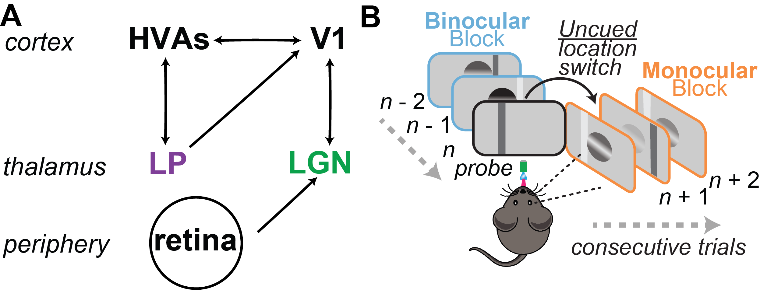
Tracking contributions of subcortical visual circuits to spatial attention and its disruption in autism spectrum disorders: People with autism spectrum disorders (ASD) often report attentional deficits that impair their ability to filter task-relevant sensory inputs from noise. It remains unclear how neural disruptions in specific brain structures could underlie impaired attention in ASD. I aim to improve our understanding of the neural basis of attentional abnormalities in ASD by tracking neural population activity in the visual thalamus of neurotypical and ASD-relevant transgenic mice performing a visual detection task that elicits spatial attention. The visual thalamus is well positioned to selectively gate the flow of visual inputs from retina to cortex depending on task demands, though it is currently unknown how single neurons of the visual thalamus contribute to attentional modulation of visual perception. Furthermore, while genetic mutations tied to ASD cause physiological disruptions to thalamic neurons, it is unknown whether or how these disruptions contribute to attentional deficits measured behaviorally. Understanding how thalamic activity is modulated with attention in neurotypical subjects is an essential step in testing whether physiological disruptions of thalamic neurons in ASD contribute to atypical sensory processing. I aim to address these questions by combining an innovative visual detection task for eliciting spatial attention in mice with high-density, multi-shank electrodes to simultaneously record from thalamic neurons with diverse functional properties.
Luminance invariant encoding in visual cortex: The visual system adapts to maintain sensitivity and selectivity over a large range of luminance intensities. One way the retina maintains sensitivity across night and day is by switching between rod and cone photoreceptors, which alters the receptive fields and interneuronal correlations of retinal ganglion cells (RGCs). While these adaptations allow the retina to transmit visual information to the brain across environmental conditions, the code used for that transmission varies. To determine how downstream targets encode visual scenes across light levels, I measured the effects of luminance adaptation on thalamic and cortical population activity. While changes in the retinal output are evident in the lateral geniculate nucleus (LGN), selectivity in primary visual cortex (V1) is largely invariant to the changes in luminance. I used a model to show that the visual system could maintain sensitivity across environmental conditions without altering cortical selectivity through the convergence of parallel functional pathways from the thalamus to cortex.

Thalamic burst and tonic firing modes are disrupted in Fragile X: Burst and tonic firing modes in the thalamus may underlie the selective and flexible relay of sensory information to cortex, a capacity which is dysregulated in Fragile X (FX) syndrome. Here we show that typical thalamic firing dynamics are disrupted in FX mutant mice. We show that a shift in the voltage sensitivity of the cellular mechanism for generating bursts contributes to reduced burst frequency and duration in FX thalamus.

ronan_oshea@utexas.edu
Contact